Introduction
Bony orbital defects can be a result of trauma, malformations and iatrogenic surgery, and can lead to many functional and cosmetic problems that often require orbit reconstruction. Numerous types of biomaterials have been used to reconstruct the orbit including autogenous materials, metallic meshes, porous polyethylene, and resorbable biomaterials (Karsloğlu et al., 2006; Potter et al., 2012; Perez et al., 2018; Gao et al., 2020). Bioresorbable implants are becoming increasingly popular as they have several attractive properties including the ability to be easily shaped, strong mechanical integrity after resorption, and do not cause complications at the donor sites (Xuan et al., 2020).
Most bone defects can be reconstructed without the need to consider bacterial infections because they are isolated from the external environment. However, grafts in the bony orbital region are vulnerable to exposure and infections. In the bony orbit, grafts are often covered by thin scar tissues adjacent to the damaged paranasal sinus mucosa that contains a high number of bacteria (Ridwan-Pramana et al., 2015; Song et al., 2016). In comparison to solid sheet materials that are easily be encapsulated, bioresorbable implants are at higher risk of infection and inflammation. Once an infection occurs, patients are at risk of different complications including exposure, migration, and the formation of fistulae and cysts.
In cases where conservative treatment is unsuccessful, orbit reconstruction may fail and require implant removal. Presoaking implants with an antibiotic solution are recommended before the placement of implant materials. However, the antibiotic effect of presoaking implants is time-limited and some antibiotics have been reported to negatively impact bone regeneration (Wu et al., 2018). To avoid infection and also promote osteogenesis, there is a critical need for the development of novel engineered bone tissue materials for orbital defect reconstruction.
Bioactive scaffold-based tissue engineering has been widely investigated and applied as a strategy for bone regeneration (Deng et al., 2013; Orciani et al., 2017; Filippi et al., 2020; Shi et al., 2020). Poly(sebacoyl diglyceride) (PSeD) with exposed hydroxyl groups is a porous bioresorbable material which can be linked to bifunctional active molecules including growth factors, peptides, and chemical groups, making it a potential scaffold for multifunctional materials (You et al., 2012; Bi et al., 2014; Wang et al., 2016; Huang et al., 2016; Gong et al., 2020).
Antibacterial peptides (ABPs) have been explored as a potential compound with antibacterial and bone regenerating properties. The first ABP named “cecropin” was found in 1980 and since then major research efforts have focused on the purification and application of ABPs (Esfandiyari et al., 2019). ABPs can kill germs in multiple innate immune system models. ABPs found in mammals can be divided into two subtypes: defensins and cathelicidins.
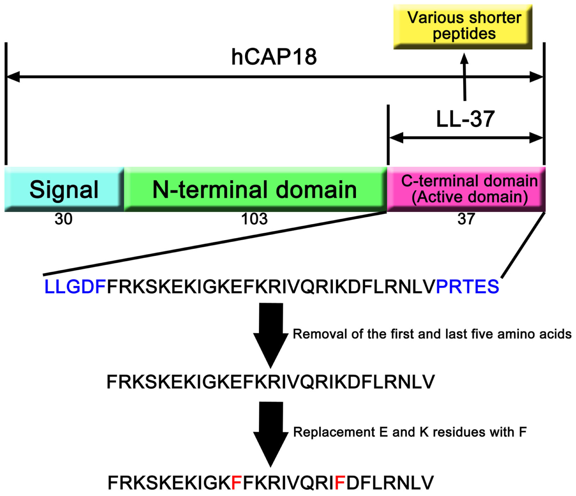
LL37 is the only known human cathelicidin antimicrobial peptide (CAMP) and is formed from 37 amino acids of the C-terminus of the human cationic antimicrobial peptide-18 (Fan et al., 2015). LL37 contributes to host defenses against intracellular infections (Stephan et al., 2016) and the prevention of infection in burn care (Vignoni et al., 2014). LL37 can also cooperate with poly (lactic-co-glycolic acid) (PLGA) to accelerate wound healing (Chereddy et al., 2014) and can be used to treat keratitis caused by bacteria (Ishida et al., 2016). LL37 has shown much potential in hematopoietic stem/progenitor cells (HSPCs) and cord blood transplantation by accelerating the process of adhesion and recruitment (Wu et al., 2012). Recently, LL-37 has been combined with titanium implants to facilitate bone formation via mesenchymal stem cell recruitment (Liu et al., 2018; Shen et al., 2019). However, the effects of LL37 osteogenesis remain to be fully understood and there is a lack of knowledge on the use of LL37 in craniofacial bone defect reconstruction.
Most of the current studies have combined LL37 with bone mesenchymal stem cells (BMSCs) which are difficult to be harvested from human bone marrow. Compared to BMSCs, human adipose-derived mesenchymal stem cells (hADSCs) can be easily collected by liposuction or doubled eyelid surgery (Mehrabani et al., 2013). Moreover, ADSCs lack HLA-ABC expression and so ADSCs are less likely to be attacked by the host immune system due to the lack of HLA-ABC expression (Menard et al., 2013). hADSCs are therefore ideal seed cells for bone regeneration and could be widely applicable to many clinical indications.
In this study, we combined the biomaterial PSeD with hADSCs and the LL37 AMP and evaluated its effects on osteogenesis in vitro using in vivo rat calvarial bone defect model. The combination technique is expected to be applied in clinical surgery.
Materials and Methods
Preparation and Characterization of PSeD Scaffolds
Poly(sebacoyl diglyceride) (PSeD) was synthesized via acid-inducedepoxide ring-opening polymerization between an equimolar amount of diglycidyl sebacate and sebacic acid in presence of 0.6 mol % tetrabutylammonium bromide in dimethylformamide at 95°C for 24 h (You et al., 2010, 2012; Huang et al., 2016). The reaction mixture was purified via precipitation in ethyl ether and vacuum-dried to yield PSeD. The porous three-dimensional scaffolds of PSeD were prepared by a modified salt fusion template method using NaCl salt particles with a size of 75–150 mM as porogen according to previous reports (Huang et al., 2016; Sun et al., 2019; Gong et al., 2020; Xuan et al., 2020).
The mechanical properties of PSeD scaffolds were evaluated via compression tests as previous description (Gong et al., 2020). The morphology of PSeD scaffolds were investigated via scanning electron microscope.
Cell Culture and Experimental Treatments
All research was approved by the Ethics Committee and the Animal Research Committee of Shanghai Ninth People’s Hospital, Shanghai Jiao Tong University School of Medicine, and informed consent was obtained from each subject. Human fat tissue was obtained from double eyelid surgery. Tissues were minced and digested with 0.1% collagenase I at 37°C with shaking at 200 rpm for 10 h. The released fat stromal cells were resuspended in Dulbecco’s modified Eagle’s medium (DMEM, Gibco) containing 10% fetal bovine serum (FBS, Gibco) and 100 units/ml of penicillin and streptomycin (Invitrogen) before incubation at 37°C in an atmosphere of 5% CO2. ADSCs were characterized by flow cytometry (CD44, CD90, CD73, CD45, CD34 and HLA-DR, all from BD Biosciences, San Jose, CA, United States), in vitro adipogenic and chondrogenic induction assays. To investigate the optimum concentrations of LL37 with the strongest osteogenic effect, after incubation for 24 h, different concentrations of LL37 (0, 1, 2, 4, 8 μg/mL) were separately added to the culture medium.
In vitro Cytotoxicity
The effects of LL37 on the proliferation of hADSCs were evaluated using a cell counting kit-8 (CCK-8) assay as previously reported (Ni et al., 2014). Cells were seeded at a density of 3.0 × 10⁴/cm2 in a 96-well plate and incubated with different concentrations of LL37 (0, 1, 2, 4, 8 μg/mL). 10 μl of CCK-8 solution was added to each well (Dojindo Molecular Technologies, Inc., Japan) incubated at 37°C for 4 h. The absorbance values at 450 nm wavelength were measured.
Detection of Osteogenic Activity in vitro
Real-Time Reverse Transcription-Polymerase Chain Reaction (RT – PCR)
After 7 days of culture, the total RNA of hADSCs was collected and purified using the EZ-press RNA purification kit (EZBioscience). Reverse transcription was performed using the PrimeScript RT reagent kit (Takara, Dalian, China). Quantitative RT-PCR was optimized with a Power SYBR Green PCR Master Mix (Applied Biosystems, Foster City, CA, United States) using a 7500 Real-Time PCR detection system. The related primer sequences are listed in Table 1 including collagen I (Col1), osteopontin (Opn), runt-related transcription factor 2 (Runx2), bone sialoprotein (BSP), and GAPDH. Relative mRNA levels were normalized to the expression level of GAPDH. All tests were performed in triplicates.
Leave a Reply